
Dr. Jessie Atterholt was invited to be a part of a special exhibit curated by Dr. Mateusz Wosik at Misericordia University, titled Lost Worlds: Microphotography of Extinct Species. The exhibit, which featured the work of an international group of early-career researchers, was curated with the goal of merging art and science by presenting the beauty of osteohistological thin sections. Within the field of vertebrate paleontology, osteohistological research has risen to prominence in recent decades as a crucial way of understanding development and life history traits in extinct organisms. The visually-striking, often colorful, thin sections produced for this research present a great opportunity for engaging the public with scientific research.
Dr. Atterholt’s submissions to the exhibit included images of thin sections from limb bones of Mirarce eatoni, a large-bodied Late Cretaceous fossil bird from Utah that she and her colleagues named and described in 2018. This specimen is highly significant for its the level of completeness (with elements from across the post-cranial skeleton represented) and incredibly detailed 3D preservation. In 2021, she published a study on the osteohistology of Mirarce, which revealed previously unknown variation in growth patterns across the skeleton. Images from this study were included in the microphotography exhibit.
Learn more about the exhibit page on the Misericordia University website.
Museum catalogue number: UCMP 139500 (University of California Museum of Paleontology)
Scientific name: Mirarce eatoni
Common name: none
Locality: Grand Escalante National Part, UT
Geological Age: Late Cretaceous
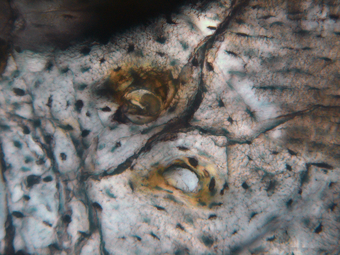
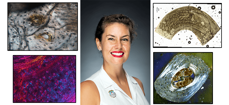
Image 1 – Humerus
The humerus of Mirarce is comprised of parallel-fibered bone with regional incipient fibrolamellar bone. This image shows developing primary osteons in a region of fibrolamellar bone, as well as fine details such as canaliculi extending from osteocyte lacunae (the tiny, squiggly arms extending from the small, black pores).
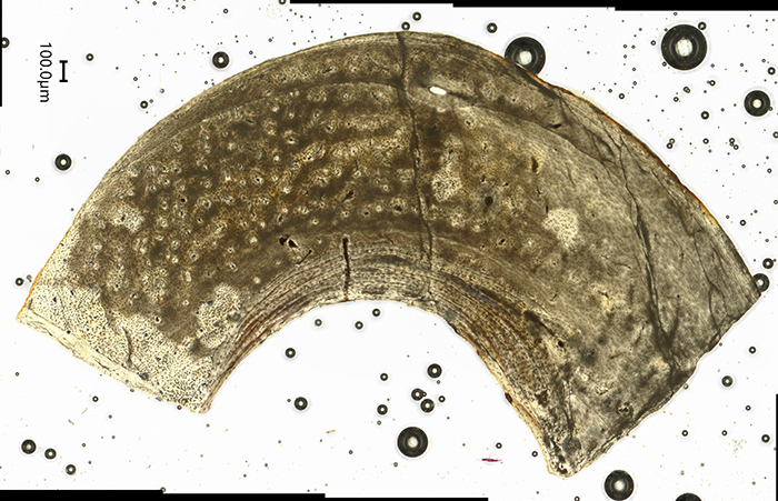
Image 2 – Metatarsal
This is a thin section through one of the tarsals of Mirarce, which is one of the long bones that makes up the foot. On the left is a large region of fast-growing fibrolamellar tissue, which grades into slower-growing parallel-fibered tissue on the right. This combination of tissue types indicates the importance of asymmetrical growth in shaping this bone as the animal grows.
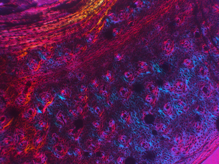
Image 3 – Metatarsal
Another image of the metatarsal bone, here shown under polarized light and zoomed in on the region of fibrolamellar bone. Here, the large, black circles are pores where blood vessels were located in the living bone, and the numerous small, black pores housed osteocytes (bone cells). This portion of the metatarsal was highly vascularized and had a high density of bone cells.
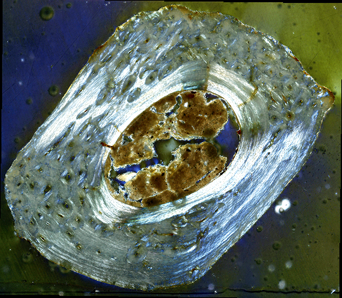
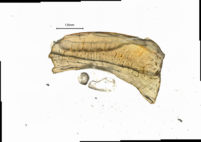
Image 4 – Phalanx
Here we see a thin section through the first phalanx (toe bone) of the second toe, shown under polarized light. This bone is primarily composed of parallel-fibered tissue, which is clearly visible in the very bright regions of the cortex. However, in the darker regions, this parallel-fibered tissue has been obscured by numerous secondary osteons. This is evidence of bone remodeling, which in this case is likely because this bone (located in the foot) was weight-bearing.
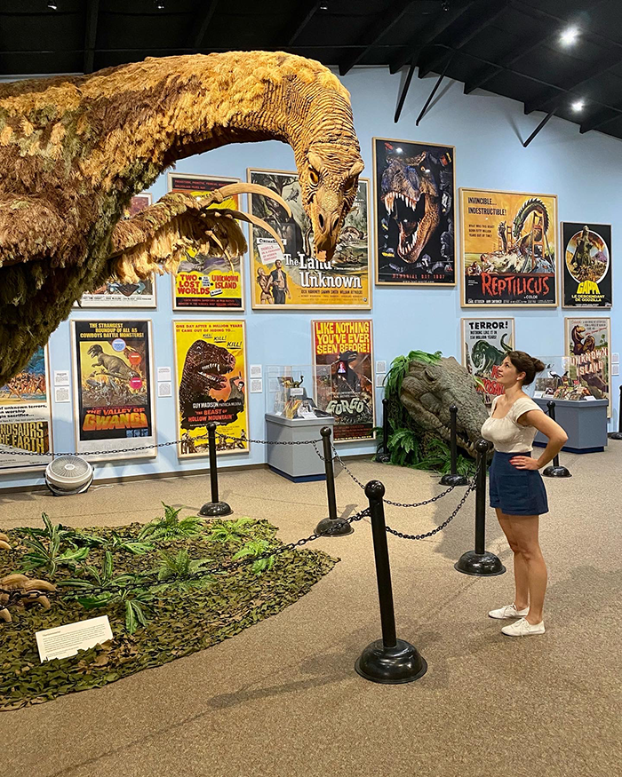
Dr. Atterholt with a reconstruction of her favorite dinosaur – the Therizinosaurus!